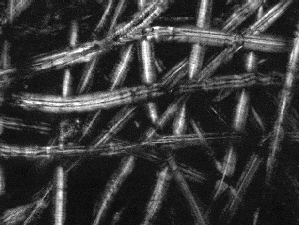
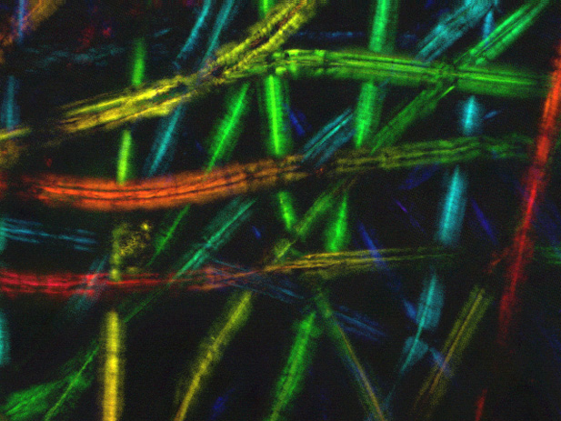
Tissue Fibers
Figure 1 - Full-Focus Composite: This tissue was imaged with an air objective under standard halogen lighting. (Scroll for additional images of this sample.)
Figure 2 - Depth Colored: Depth information has been added to the image above through color with red corresponding to the higher points and blue the lower. The color further separates the individual fibers. (Scroll for additional images of this sample.)