| Products | Who we are | Employment | Contact Us | Home |
|
|
|
||||
Image Gallery |
Optical Devices |
Software |
Confocal Equipment |
||
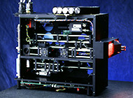
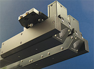
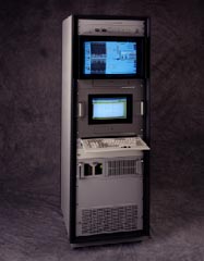
Triptar designs, builds, and maintains full turnkey optical instruments. Here are some details about two instrument projects.
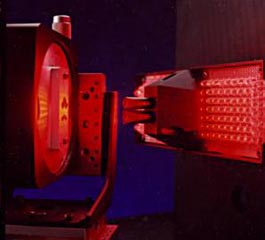
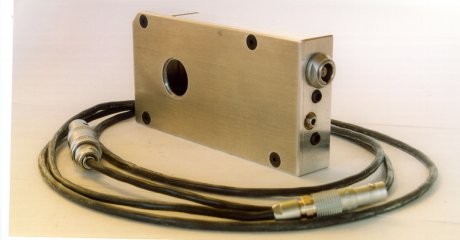
 Fully automated xyz-theta measurements through a custom designed dual-field microscope. Features xy measurement of geometric features in the fields of view; binary zoom system for detailed measurement mode or general navigational mode; Z distance sensing along the line of sight; and angular tilt of the specimen with an integrated autocollimator.
 sized(200x110).jpg)
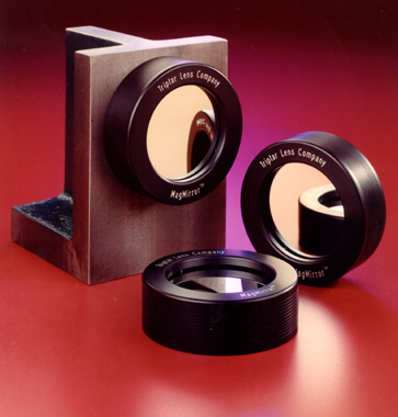
Triptar designed the first commercially available structured light confocal microscope, Optigrid™, on the basis of patents developed at Oxford University. Triptar designed the opto-mechanical module that replaces the field stop in the illuminator of a standard brightfield microscope, the amplifier to activate the piezo-translated pattern generator, and the machine control software to coordinate the translation of the pattern with image acquisition and movement of the specimen stage. Triptar also designed the user interface by combining proprietary and off-the-shelf software such as Media Cybernetics' Image-Pro™. The multi-year product development project was conducted on behalf of Avimo Precision Instruments, which owned the exclusive, global rights to the patents and the Optigrid trademark. Triptar served as the premier dealer and established and supported the first network of dealers in North America, Europe, and Asia.

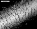
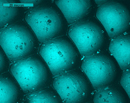
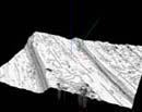
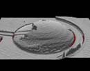
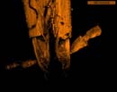
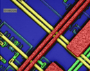
A confocal microscope presents only the plane of focus to the eye or to the camera. Out of focus planes are suppressed. This performance is sometimes called "optical sectioning" because it mimics the technique of slicing a specimen into thin planes to observe its internal 3-dimensional structures.
After collecting a set of thin optical sections, the 3D structure of the specimen may be displayed after image processing by cycling through the sections, rendering the sections as a solid body viewed at different angles, or by presenting a stereo pair for direct observation. Sometimes, a confocal microscope is used only to produce enhanced contrast images because the isolated image plane is not confounded by out of focus detail. The entire stack of optical sections can be combined into one high contrast image with all levels in sharp focus.
Most of the images in the Image Gallery were acquired with SurfaceScope™, a special Triptar version of Optigrid™, and rendered with Triptar's image reconstruction software, SurfaceView™. The images showcase the remarkable depth and breadth of this confocal technology.
|
||||||||
|
All images Copyright ©1993-2015 Triptar Lens Company, Inc. |
||||||||